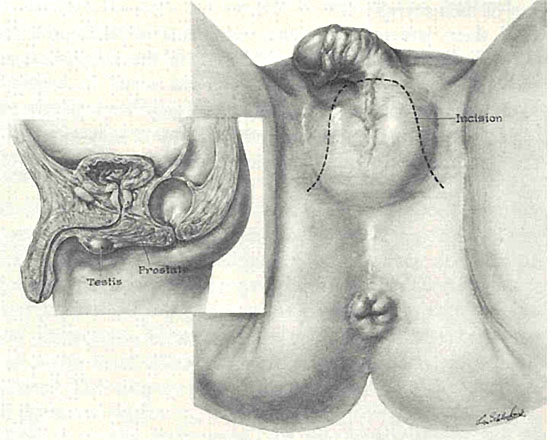
Figure 1. A sketch of the perineum showing the line of primary incision.
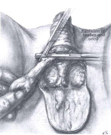
Figure 2. The right spermatic cord is clamped and ligated.
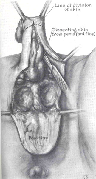
Figure 3. The primary incision is continued up the ventral side of the shaft of the penis.
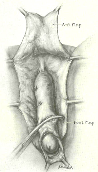
Figure 4. The anterior flap is developed from the skin of the penis.
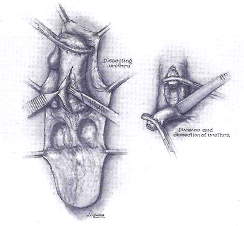
Figure 5. The urethra is dissected from the shaft of the penis.
Figure 6. The corpora cavernosa are separated to assure a minimal stump.
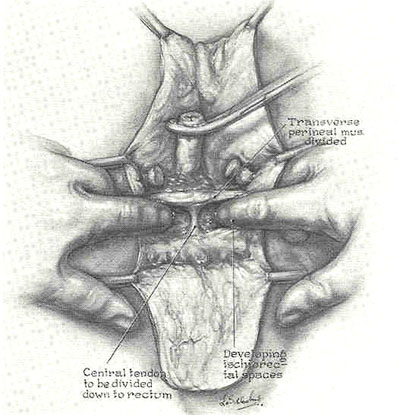
Figure 7. The perineal dissection.
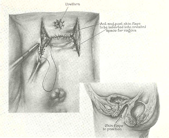
Figure 8. The perineal dissection has been completed and the anterior flap perforated to position the urethral meatus.
Figure 9. The skin flaps are sutured and placed in position in the vaginal cavity.
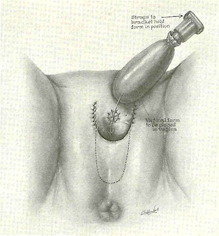
Figure 10. The preservation of the vaginal cavity is assured by use of a suitable vaginal form.
Note 1: Figure 10 is quite misleading and does not correspond to the anatomy the should result from this procedure. In figure 10, the vaginal opening is way too far forward from the anal opening, and the vaginal entry is shown going first in horizontally and then turning upwards after passing a large web of skin in front of the anus. (Compare this sketch with the later photos of the details of modern SRS results, especially the one showing the entry of a vaginal stent into a postop's vagina). This very poorly conceived sketch has likely been the source of many botched surgeries in the early days, as surgeons copying the Hopkins procedure may have thought that a thick web of skin was needed in order to prevent tears into the rectum. Such webs of skin often prevented easy dilations and intercourse for patients after SRS, leading to vaginal stenosis (loss of depth and/or width).
Note 2: Over the years, the techniques for doing SRS have been steadily refined. It has also became common for post-op MtF's to have additional genital surgery called "labiaplasty" that construct further details of the external female genitalia. For more information on modern SRS surgeries, see the links and the "Photo Details of Modern SRS Results" below.


No comments:
Post a Comment